Brain Tissue Segmentation Using NeuroNet With Different Pre-processing Techniques
 Used architecture
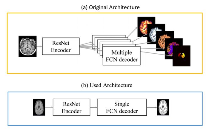
Abstract
Automatic segmentation of brain Magnetic Resonance Imaging (MRI) images is one of the vital steps for quantitative analysis of brain for further inspection. In this paper, NeuroNet has been adopted to segment the brain tissues (white matter (WM), grey matter (GM) and cerebrospinal fluid (CSF)) which uses Residual Network (ResNet) in encoder and Fully Convolution Network (FCN) in the decoder. To achieve the best performance, various hyper-parameters have been tuned, while, network parameters (kernel and bias) were initialized using the NeuroNet pre-trained model. Different pre-processing pipelines have also been introduced to get a robust trained model. The model has been trained and tested on IBSR18 data-set. To validate the research outcome, performance was measured quantitatively using Dice Similarity Coefficient (DSC) and is reported on average as 0.84 for CSF, 0.94 for GM, and 0.94 for WM. The outcome of the research indicates that for the IBSR18 data-set, pre-processing and proper tuning of hyper-parameters for NeuroNet model have improvement in DSC for the brain tissue segmentation.