Fakrul Islam tushar is a Medical Imaging & Computer Vision Engineer, primarily engaged in research, computer-aided diagnosis and healthcare innovation using machine learning, neural networks, and image analysis driven solutions. He graduated from the Erasmus+ Joint Master in Medical Imaging and Applications and is currently a Post-Graduate Research Associate at the Carl E. Ravin Advanced Imaging Laboratories (RAI Labs), Duke University Medical Center, USA. He earned a Bachelor of Science degree in Electrical and Electronics Engineering at American International University Bangladesh (AIUB). He is a recipient of the “Cum Laude” distinction and academic honor at the 17th Convocation Ceremony of AIUB, “Dean’s Award” for his final-year undergraduate research project, the European Union: Erasmus+ Grant and the Duke University: Masters Thesis Grant. His past affiliations include IEEE AIUB Student Branch, Teach For Bangladesh and “Literacy Through Leadership”.
Associate in Research, 2019-present
Duke University Medical Center
Masters in Medical Imaging and Applications, 2017-2019
Erasmus Mundus Joint Master ( University of Burgundy; University of Cassino; University of Girona, Duke University)
BSc in Electrical & Electronic Engineering, 2013-2017
American International University Bangladesh
Automatic segmentation of brain Magnetic Resonance Imaging (MRI) images is one of the vital steps for quantitative analysis of brain for further inspection. In this paper, NeuroNet has been adopted to segment the brain tissues (white matter (WM), grey matter (GM) and cerebrospinal fluid (CSF)) which uses Residual Network (ResNet) in encoder and Fully Convolution Network (FCN) in the decoder. To achieve the best performance, various hyper-parameters have been tuned, while, network parameters (kernel and bias) were initialized using the NeuroNet pre-trained model. Different pre-processing pipelines have also been introduced to get a robust trained model. The model has been trained and tested on IBSR18 data-set. To validate the research outcome, performance was measured quantitatively using Dice Similarity Coefficient (DSC) and is reported on average as 0.84 for CSF, 0.94 for GM, and 0.94 for WM. The outcome of the research indicates that for the IBSR18 data-set, pre-processing and proper tuning of hyper-parameters for NeuroNet model have improvement in DSC for the brain tissue segmentation.
The segmentation is an essential part of many computer vision systems and medical applications. The goal is to divide an input image into a set of non-overlapping regions which union is the entire image. The Expectation-Maximization (EM) algorithm is an iterative technique designed for probabilistic models. Expectation-Maximization algorithm for segmenting brain MRI images (T1-w image) into the three main tissues: white matter (WM), grey matter (GM) and cerebrospinal fluid (CSF).
The idea of the project is to apply 2D U-net to Segment the RIO using the publicly available data from ISIC Challenge 2017 and classification of the segmented region for classification of lesion. Multiple OTS model ( Resnet, VGG, Densenet) were used for classification. Among the model VGG out performed all other model. Ensembling of the models was also applied and performed better than single classifier, Democracy always wins.
Lung diseases classification in 2D using chest CT cases and Analysis the multi-channel effect on classification. This work is been done during summer internship July-Aguest 2018, Duke University Medical Center.
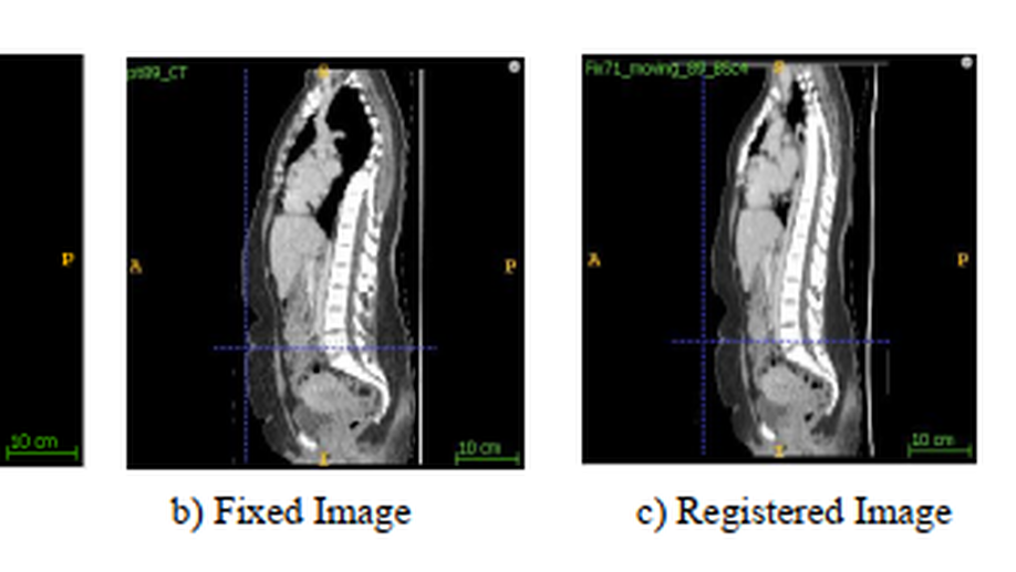
Main idea of this project was making some new data using simple registration technique from existing CT data. This code be a possible alternative of arbitrary augmentation such as flip, rotation, zoom etc. A simple but powerful non-rigid registration technique BSpline is been used for this. There are number of parameters that can be used to make the variation of the registration to make new data which is completely different from Moving and Fixed CT used for registration.
Skin lesion segmentation is one of the first steps towards automatic Computer-Aided Diagnosis of skin cancer. Vast variety in the appearance of the skin lesion makes this task very challenging. The contribution of this paper is to apply a power foreground extraction technique called GrabCut for automatic skin lesion segmentation with minimal human interaction in HSV color space. Preprocessing was performed for removing the outer black border. Jaccard Index was measured to evaluate the performance of the segmentation method. On average, 0.71 Jaccard Index was achieved on 1000 images from ISIC challenge 2017 Training Dataset.
s the learning rate is one of the most importanthyper-parameters to tune for training convolutional neural net-works. In this paper, a powerful technique to select a range oflearning rates for a neural network that named cyclical learningrate was implemented with two different skewness degrees. Itis an approach to adjust where the value is cycled between alower bound and upper bound. CLR policies are computationallysimpler and can avoid the computational expense of fine tuningwith fixed learning rate. It is clearly shown that changing thelearning rate during the training phase provides by far betterresults than fixed values with similar or even smaller number of epochs.
Mammography is the most widely used gold standard method for the screening of the breast cancer and Mass detection is the prominent pre-processing step. State-of-art performances of the DCNN architectures in the field of classification made them an obvious choice for the image classifications. However, due to limited availability of medical data, application of DCNN architectures in medical images is challenging. In this project our contribution was mass detection in mammographic images using two different approaches. First approach was applying transfer learning concept which lessen the demand of data to use a pre-trained publicly available VGG16 model and second approach was training a Alexnet from the scratch to classify mass and non-mass mammographic images. Both the models were trained and tested with different hyperparameters and different data size. From the experiment results, it can be concluded that transfer learning approach for VGG16, training from fully connected layer 2 (fc2) has promising expected result for mass detection. In our research, maximum accuracy for mass detection was 93.60% for VGG16 with 13500 train images.
Design an inverse kinematic controller to move end-effector. This work is done as a coursework for the 2nd semester of MAIA in Introduction to Robotics course.The task was to design an Inverse Kinematic controller to move the end-effector from the position pA to the position pB The movement should be repeated 3 times by control: 1)the position-only without exploiting the redundancy 2)the position and the orientation without exploiting the redundancy (desired orientation equal to the initial one) 3)the position and the orientation avoiding an obstacle put in p0 (desired orientation equal to the initial one)
Left ventricular non-compaction (LVNC) is a rare cardiomyopathy (CMP) that should be considered as a possible diagnosis because of its potential complications which are heart failure, ventricular arrhythmias, and embolic events. For analysis cardiac functionality, extracting information from the Left ventricular (LV) is already a broad field of Medical Imaging. Different algorithms and strategies ranging that is semiautomated or automated has already been developed to get useful information from such a critical structure of heart. Trabeculae in the heart undergoes difference changes like solid from spongy. Due to failure of this process left ventricle non-compaction occurred. In this project, we will demonstrate the fractal dimension (FD) and manual segmentation of the Magnetic Resonance Imaging (MRI) of the heart that quantify amount of trabeculae inside the heart. The greater the value of fractal dimension inside the heart indicates the greater complex pattern of the trabeculae in the heart.
The availability of sophisticated source attribution techniques raises new concerns about privacy and anonymity of photographers, activists, and human right defenders who need to stay anonymous while spreading their images and videos. An image can be considered to be a combination of both significant (foreground) objects and some less significant (background) objects. Content aware image resizing (CAIR) algorithm uses the different edge detection methods to segregate the useful objects from the background. When applied to an image, CAIR can resize the image to a very different aspect ratio without destroying the aspect ratio of the useful objects in the image. In this project, we simply implement a content aware image resizing (CAIR) in MATLAB environment. The main idea to implement CAIR is to remove or insert the vertical or horizontal seams (paths of pixel) having the lowest energy. After implanted the Seam Carving Algorithm for Content aware image resizing (CAIR), analysis shows that the implemented seam carving for CAIR can generate more desirable resized images than cropping, resampling, and conventional seam carving techniques.
Few years back, face recognition is only limited to personal identification, now this technology is been grown in the field of security, healthcare, personalized services etc. Many methods are developed to perform for face recognition and they work quit well.The basic idea of this project it to compute a data set of features from the face images 𝐼𝑞which have compared with a set of database images 𝐼1………𝐼𝑞 to detect the best match. Every pixel can be considered as a variable that means the dimensional space is very high. PCA is been used to decreasing the dimensionality of the image for making this recognition problem easier.
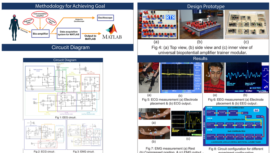
This paper presents the design, development and implementation of a reconfigurable low-cost biopotential amplifier trainer module (RTR module) and quantitative analysis of the students’ compatibility with the trainer module. The trainer module can measure Electrocardiogram (ECG), Electroencephalogram (EEG) and Electromyogram (EMG) biopotential signals by reconfiguring the module using the basic circuit and filtering blocks. Given hand on experience, the module is designed and implemented in such reconfigurable manner that the students can avoid, disconnect and add any filtering blocks to understand the effect of these filters to the biopotential signals. The laboratory experience is an important component of the learning process. The RTR module is a low cost and compact educational tool. With this RTR module, the students should be able to recognize the biopotential signals and the acquisition methods in an intuitive and easy way, allowing them to improve their skills of designing biomedical instrumentation.
The name of our Application is ASHA Which mean Hope in Bengali. Here ASHA Stands for A Solution to Help Autism. The Idea of our App ASHA is to introduce a detection or test procedure for Autism.Autism is a spectrum of closely related disorders with a shared core of symptoms. Autism spectrum disorders appear in infancy and early childhood, causing delays in many basic areas of development, such as learning to talk, play, and interact with others.
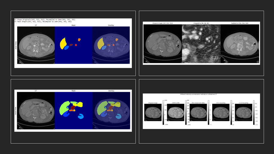
In this blog, I will introduce a Library for 3D augmentations called volumentations-3D. .
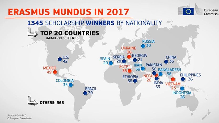
Trying cover the FAQs abour ERASMUS+ Scholarship funed By European Union.
Automatic segmentation of skin lesions is considered a crucial step in Computer-aided Diagnosis (CAD) systems for melanoma detection. Despite its significance, skin lesion segmentation remains an unsolved challenge due to their variability in color, texture, and shapes and indistinguishable boundaries.Through this study, we present a new and automatic semantic segmentation network for robust skin lesion segmentation named Dermoscopic Skin Network (DSNet). In order to reduce the number of parameters to make the network lightweight, we used a depth-wise separable convolution in lieu of standard convolution to project the learned discriminating features onto the pixel space at different stages of the encoder. Additionally, we implemented both a U-Net and a Fully Convolutional Network (FCN8s) to compare against the proposed DSNet. We evaluate our proposed model on two publicly available datasets, namely ISIC-20171 and PH22 . The obtained mean Intersection over Union (mIoU) is 77.5% and 87.0% respectively for ISIC-2017 and PH2 datasets which outperformed the ISIC-2017 challenge winner by 1.0% with respect to mIoU. Our proposed network also outperformed U-Net and FCN8s respectively by 3.6% and 6.8% with respect to mIoU on the ISIC-2017 dataset. Our network for skin lesion segmentation outperforms the other methods discussed in the article and is able to provide better-segmented masks on two different test datasets which can lead to better performance in melanoma detection. Our trained model along with the source code and predicted masks are made publicly available
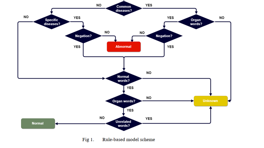
Lack of annotated data is a major challenge to machine learning algorithms, particularly in the field of radiology. Algorithms that can efficiently extract labels in a fast and precise manner are in high demand. Weak supervision is a compromise solution, particularly, when dealing with imaging modalities like Computed Tomography (CT), where the number of slices can reach 1000 per case. Radiology reports store crucial information about clinicians’ findings and observations in CT slices. Automatic generation of labels from CT reports is not a trivial task due to the complexity of sentences and diversity of expression in free-text narration. In this study, we focus on abnormality classification in lungs, liver and kidneys. Firstly, a rule-based model is used to extract weak labels at the case level. Afterwards, attention guided recurrent neural network (RNN) is trained to perform binary classification of radiology reports in terms of whether the organ is normal or abnormal. Additionally, a multi-label RNN with attention mechanism is trained to perform binary classification by aggregating its output for four representative diseases (lungs: emphysema, mass-nodule, effusion and atelectasis-pneumonia; liver: dilatation, fatty infiltration-steatosis, calcification-stone-gallstone, lesion-mass; kidneys: atrophy, cyst, stone-calculi, lesion) into a single abnormal class. Performance has been evaluated using the receiver operating characteristic (ROC) area under the curve (AUC) on 274, 306 and 278 reports for lungs, liver and kidneys correspondingly, manually annotated by radiology experts. The change in performance was evaluated for different sizes of training dataset for lungs. The AUCs of multi-label pretrained models: lungs - 0.929, liver - 0.840, kidney - 0.844; multi-label models: lungs - 0.903, liver - 0.848, kidney - 0.906; binary pretrained models: lungs - 0.922, liver - 0.826, kidneys - 0.928.
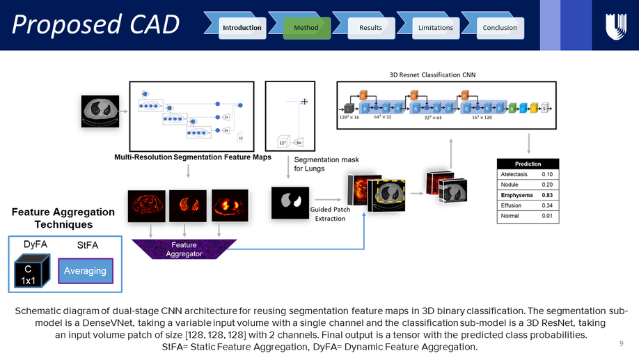
Weakly supervised disease classification of CT imaging suffers from poor localization owing to case-level annotations, where even a positive scan can hold hundreds to thousands of negative slices along multiple planes. Furthermore, although deep learning segmentation and classification models extract distinctly unique combinations of anatomical features from the same target class(es), they are typically seen as two independent processes in a computer-aided diagnosis (CAD) pipeline, with little to no feature reuse. In this research, we propose a medical classifier that leverages the semantic structural concepts learned via multi-resolution segmentation feature maps, to guide weakly supervised 3D classification of chest CT volumes. Additionally, a comparative analysis is drawn across two different types of feature aggregation to explore the vast possibilities surrounding feature fusion. Using a dataset of 1593 scans labeled on a case-level basis via rule-based model, we train a dual-stage convolutional neural network (CNN) to perform organ segmentation and binary classification of four representative diseases (emphysema, pneumonia/atelectasis, mass and nodules) in lungs. The baseline model, with separate stages for segmentation and classification, results in AUC of 0.791. Using identical hyperparameters, the connected architecture using static and dynamic feature aggregation improves performance to AUC of 0.832 and 0.851, respectively. This study advances the field in two key ways. First, case-level report data is used to weakly supervise a 3D CT classifier of multiple, simultaneous diseases for an organ. Second, segmentation and classification models are connected with two different feature aggregation strategies to enhance the classification performance.
Automatic segmentation of brain Magnetic Resonance Imaging (MRI) images is one of the vital steps for quantitative analysis of brain for further inspection. In this paper, NeuroNet has been adopted to segment the brain tissues (white matter (WM), grey matter (GM) and cerebrospinal fluid (CSF)) which uses Residual Network (ResNet) in encoder and Fully Convolution Network (FCN) in the decoder. To achieve the best performance, various hyper-parameters have been tuned, while, network parameters (kernel and bias) were initialized using the NeuroNet pre-trained model. Different pre-processing pipelines have also been introduced to get a robust trained model. The model has been trained and tested on IBSR18 data-set. To validate the research outcome, performance was measured quantitatively using Dice Similarity Coefficient (DSC) and is reported on average as 0.84 for CSF, 0.94 for GM, and 0.94 for WM. The outcome of the research indicates that for the IBSR18 data-set, pre-processing and proper tuning of hyper-parameters for NeuroNet model have improvement in DSC for the brain tissue segmentation.
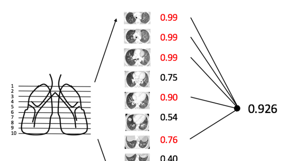
Our goal is to investigate using only case-level labels extracted automatically from radiology reports to construct a multi-disease classifier for CT scans with deep learning method. We chose four lung diseases as a start: atelectasis, pulmonary edema, nodule and pneumonia. From a dataset of approximately 5,000 chest CT cases from our institution, we used a rule-based model to analyze those radiologist reports, labeling disease by text mining to identify cases with those diseases. From those results, we randomly selected the following mix of cases: 275 normal, 170 atelectasis, 175 nodule, 195 pulmonary edema, and 208 pneumonia. As a key feature of this study, each chest CT scan was represented by only 10 axial slices (taken at regular intervals through the lungs), and furthermore all slices shared the same label based on the radiology report. So the label was weak, because often disease will not appear in all slices. We used ResNet-50[1] as our classification model, with 4-fold cross-validation. Each slice was analyzed separately to yield a slice-level performance. For each case, we chose the 5 slices with highest probability and used their mean probability as the final patient-level probability. Performance was evaluated using the receiver operating characteristic (ROC) area under the curve (AUC). For the 4 diseases separately, the slice-based AUCs were 0.71 for nodule, 0.79 for atelectasis, 0.96 for edema, and 0.90 for pneumonia. The patient-based AUC were 0.74 for nodule, 0.83 for atelectasis, 0.97 for edema, and 0.91 for pneumonia. We backprojected the activations of last convolution layer and the weights from prediction layer to synthesize a heat map [2] . This heat map could be an approximate disease detector, also could tell us feature patterns which ResNet-50 focus on
This paper presents the design, development and implementation of a reconfigurable low-cost biopotential amplifier trainer module (RTR module) and quantitative analysis of the students’ compatibility with the trainer module. The trainer module can measure Electrocardiogram (ECG), Electroencephalogram (EEG) and Electromyogram (EMG) biopotential signals by reconfiguring the module using the basic circuit and filtering blocks. Given hand on experience, the module is designed and implemented in such reconfigurable manner that the students can avoid, disconnect and add any filtering blocks to understand the effect of these filters to the biopotential signals. The laboratory experience is an important component of the learning process. The RTR module is a low cost and compact educational tool. With this RTR module, the students should be able to recognize the biopotential signals and the acquisition methods in an intuitive and easy way, allowing them to improve their skills of designing biomedical instrumentation.